
A Clue to How Dementia Related Damage Spreads
South Korean researchers have identified a protein called FAM84B that helps tau move between brain cells, revealing a possible link between inflammation in the brain and a process associated with the progression of Alzheimer's disease. In cell experiments, increasing FAM84B caused more tau to leave nerve cells and reach neighboring cells. A drug that blocked related calcium signaling reduced that transfer under inflammatory conditions.
Contents
- A Clue to How Dementia Related Damage Spreads
- What Researchers Found in Human Samples
- Why Tau Moving Between Cells Matters
- Inflammation Connects to the Transfer Process
- How Calcium Helps Tau Leave a Cell
- What Happened When Researchers Used Dantrolene?
- A Research Lead in a Country Facing Rising Care Needs
- From Publication to the Next Experiments
- Key Points
The Korea National Institute of Health, part of the Korea Disease Control and Prevention Agency, announced the findings on November 4, 2026. The research was published online on October 2, 2026, in the Journal of Neuroinflammation. Its central finding concerns how cells release and receive tau, a protein that normally helps maintain the internal structure of nerve cells but can damage them when it changes abnormally and accumulates.
The researchers combined observations from human brain tissue with experiments using nerve cells made from patient stem cells. Their work points to a sequence in which inflammatory signals increase FAM84B, calcium signaling helps cells release tau, and nearby cells take it up. The next research priority is to test how this pathway affects brain damage, disease progression and cognitive function in animals.
What Researchers Found in Human Samples
The team's Journal of Neuroinflammation study identifies FAM84B, also known as LRATD2, as a regulator of tau transfer between cells. Researchers found higher amounts of the protein in postmortem brain tissue from people with Alzheimer's disease, progressive supranuclear palsy and primary age-related tauopathy.
These conditions belong to a group called tauopathies, diseases involving abnormal tau accumulation in the brain. Finding increased FAM84B across several such conditions gave the researchers a reason to investigate its role beyond a single diagnosis.
Higher FAM84B levels also appeared in cortical nerve cells made from stem cells derived from people with Alzheimer's disease, compared with cells from people without dementia. Cortical cells come from the brain's outer region, which supports functions including memory and thinking. Using patient tissue alongside living cells allowed the team to investigate both disease associated patterns and the cellular processes behind them.
Why Tau Moving Between Cells Matters
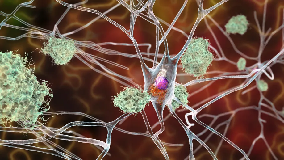
Tau normally binds to microtubules, tiny structures that help support the shape and internal organization of nerve cells. When tau becomes abnormal and builds up, it can contribute to nerve cell damage. Alzheimer's disease involves both abnormal tau and the accumulation of another substance, amyloid beta.
Tau spreading to neighboring cells is associated with disease progression. Researchers therefore want to understand more than how much tau is present inside a cell. They also need to know how it gets out, what controls that release and how another cell receives it.
FAM84B appears to influence that movement. When researchers increased its amount in nerve cells, tau inside those cells decreased while tau released outside them increased. The number of neighboring cells receiving tau also rose. Taken together, these measurements describe a shift in tau's location, from within the original cells toward the surrounding space and other cells.
The paper places this work within growing interest in tau as a treatment target. Its authors note that the modest clinical benefits achieved through research focused on amyloid beta have encouraged investigation of other processes involved in Alzheimer's disease, including the movement of tau.
Inflammation Connects to the Transfer Process
The researchers exposed nerve cells to interleukin-6 and tumor necrosis factor alpha, commonly shortened to IL-6 and TNF-alpha. Both are cytokines, chemical messages used by cells during inflammatory responses. Under these experimental conditions, FAM84B expression increased and more tau moved between cells.
The study also associated activation of STAT3 with increased tau transfer. STAT3 is a protein that helps carry signals inside a cell and influences gene activity. Its involvement adds another part to the proposed connection between inflammatory messages and changes in how nerve cells handle tau.
Cho Chul-man, the senior researcher at the Korea National Institute of Health who led the study, described the finding's contribution:
This study is the first to show that FAM84B plays an important role in the process through which neuroinflammation affects tau transmission.
The experiments give researchers a specific pathway to investigate: inflammatory signals, increased FAM84B and greater movement of tau. That is more precise than observing inflammation and abnormal tau in the same brain, because the cell experiments allowed the researchers to change conditions and measure the resulting transfer.
How Calcium Helps Tau Leave a Cell
RYR3, short for ryanodine receptor 3, played a major role in the tau release process described by the researchers. Ryanodine receptors help control the release of calcium from stores inside cells. Calcium acts as a signal that can trigger cellular activities, including the release of material.
The paper links increased FAM84B to tau secretion associated with RYR3 calcium signaling. Its title describes this as exocytic tau export. Exocytosis is a process through which a cell releases material outside its boundary.
Neighboring cells then took up the released tau partly through clathrin associated endocytosis. Endocytosis means bringing material into a cell. Clathrin is a protein involved in forming small packages that help carry material inward from the cell surface.
The results therefore describe two connected stages: tau leaving one cell and entering another. Clathrin associated uptake accounted for part of the receiving process, while RYR3 linked calcium signaling was involved in release. Keeping those stages separate helps explain why researchers measured both the amount of tau outside cells and the number of cells that received it.
What Happened When Researchers Used Dantrolene?
The team tested whether blocking ryanodine receptor signaling could reduce tau transfer. They treated nerve cells exposed to inflammatory stimuli with dantrolene, a drug that inhibits these receptors.
Dantrolene suppressed the associated calcium signaling and reduced the number of neighboring cells receiving tau. This gives researchers an experimental way to intervene in the pathway and study how changes in calcium signaling affect tau movement.
The drug experiment targeted receptor signaling within the pathway. FAM84B remains the protein the researchers identified as helping connect inflammation to increased tau release and transfer. Future work will need to determine which parts of that process could be useful treatment targets.
Cho said the next stage must address disease outcomes:
Further research is needed to determine how this pathway affects actual disease progression and assess its potential as a therapeutic target.
Animal studies are needed to examine the spread of tau pathology, nerve cell injury and cognitive function. Those outcomes will help determine whether reducing transfer through this pathway can protect the brain.
A Research Lead in a Country Facing Rising Care Needs
South Korea's dementia burden gives the research a concrete public health context. An estimated 9.2% of people aged 65 and older had dementia in 2023, amounting to about 870,000 people. That share is roughly one person in every 11 in that age group.
Dementia caused 14,251 deaths in South Korea that year. Annual national dementia care costs were estimated at 22.9 trillion won. The figures describe the scale of illness and care needs that motivate research into how dementia begins and progresses.
Kim Won-ho, director of the institute's department of chronic disease convergence research, said understanding the biological mechanisms behind disease onset and progression is essential to addressing dementia. He said the institute plans to connect research using human biological resources with studies based on stem cells to build evidence for further investigation.
The FAM84B project follows that approach. Postmortem tissue provides information about changes present in human disease, while patient derived nerve cells allow researchers to manipulate a possible mechanism in the laboratory. The study received funding through Korea Disease Control and Prevention Agency grants 2022-NG-008-02, 2025-NI-005-01 and 2023-NS-001-00.
From Publication to the Next Experiments
The findings reached the scientific journal before the institute's public announcement. The key dates and next research steps are:
- 2023: South Korea's estimated dementia prevalence among people aged 65 and older was 9.2%, representing about 870,000 people.
- October 2, 2026: The FAM84B study was published online in the Journal of Neuroinflammation.
- November 4, 2026: The Korea National Institute of Health announced the findings.
- Next research stage: Animal experiments are needed to test effects on tau pathology, nerve cell damage and cognition.
For people living with dementia and their families, the immediate development is a more detailed biological explanation to guide treatment research. The practical question now is whether interrupting this process can slow the damage that affects memory, thinking and daily life. No date was given for results from the proposed follow-up work.
Key Points
- Researchers found increased FAM84B in brain tissue from people with several tau related conditions and in nerve cells derived from Alzheimer's patients.
- Increasing FAM84B in cell experiments increased tau release and the number of neighboring cells receiving it.
- Inflammatory signals increased FAM84B expression and tau transfer, with STAT3 activation also associated with increased transfer.
- RYR3 linked calcium signaling helped drive release, while clathrin associated uptake contributed to tau entering neighboring cells.
- Dantrolene reduced transfer in laboratory cells exposed to inflammatory stimuli.
- The next priority is testing the pathway's effects on disease progression, brain injury and cognition in animals.