
A few millimeters could change a child's treatment
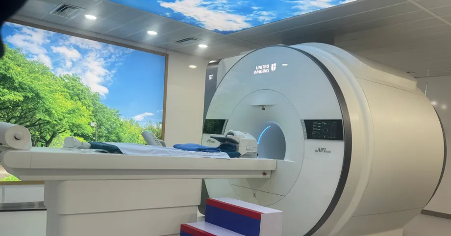
Hanoi Medical University Hospital put a 5 Tesla whole body MRI scanner into operation on the morning of October 26, introducing what hospital leaders and Vietnam's radiology association describe as the first system of its kind in Southeast Asia. Its purpose is especially concrete in difficult cases: finding abnormalities so small that doctors struggle to identify them with existing equipment.
Contents
- A few millimeters could change a child's treatment
- What does 5 Tesla mean?
- Why the brain is an early priority
- Following blood flow without injected contrast
- Looking at the chemistry of tumors
- A whole body system with wider research uses
- Training began before the launch
- Building a research program around Vietnamese patients
- Safety, cost and results will shape access
- Key Points
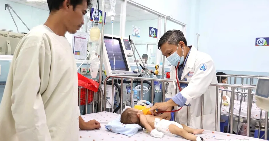
Hospital director Associate Professor Nguyen Lan Hieu described an eight year old child with recurrent epilepsy that had resisted treatment. A scan using a 3T machine showed a suspected abnormality in the cerebral cortex, the brain's outer layer. Images from the new 5T system helped identify a focus of cortical dysplasia measuring only a few millimeters.
Cortical dysplasia means an area of the brain's outer layer developed abnormally. Such an area can be involved in seizures. Locating it more precisely gives doctors information they need when considering treatment for a child whose seizures continue despite medication.
The scanner, the uMR Jupiter made by United Imaging Healthcare, is intended for both patient care and research. Hieu said its value would depend on trained clinicians using it for appropriate cases. The hospital has committed to judging the investment through better diagnosis, scientific work and specialist training rather than the number of scans performed.
What does 5 Tesla mean?
Tesla is the unit used to describe the strength of an MRI scanner's magnetic field. MRI uses a magnetic field and radio waves to produce images of structures inside the body. The 1.5T and 3T systems already widely used in hospitals remain the comparison points for the new machine.
A field of 5T is about 67 percent stronger than 3T, based on their stated field strengths. The practical benefit sought in Hanoi is more useful imaging information: finer detail and clearer differences between tissues.
Hieu explained the technical advantage at the launch, describing how the scanner separates useful information from unwanted interference in an image:
The Jupiter 5T uMR system uses a 5 Tesla magnetic field, capable of generating images with a higher signal-to-noise ratio and tissue contrast compared to currently popular 1.5T and 3T magnetic resonance imaging systems. This feature opens up the possibility of more detailed examination of microstructures and functions of many organs in the body.
In simple terms, a higher signal compared with noise can make small features easier to see. Better tissue contrast helps doctors distinguish neighboring structures that might otherwise look similar.
Why the brain is an early priority
Professor Pham Minh Thong, chair of the Vietnam Association of Radiology and Nuclear Medicine, said 5T systems remain rare worldwide. He identified the Hanoi installation as Southeast Asia's first and said the technology could support more detailed study of organ structure and function.
Neurology is a major focus. Beyond the child's epilepsy case, the hospital is interested in examining small brain structures, blood vessel walls and changes associated with Parkinson's disease and other neurological conditions.
Detailed images of the hippocampus, a brain structure involved in memory, may help doctors investigate certain forms of epilepsy. Research applications also include examining changes around small blood vessels and structures relevant to neurodegenerative disease.
Hieu stressed the importance of assessing function as well as anatomy. Anatomy shows what an organ looks like; functional imaging provides information about how its tissues are working. For the brain, that can include the movement of blood through tissue. Similar questions apply to the heart and liver.
Following blood flow without injected contrast
The hospital also plans to explore techniques that examine brain blood vessels and perfusion without an injected contrast agent. Perfusion means the delivery of blood to tissue, which supplies the oxygen and nutrients that tissue needs.
One technique discussed in connection with the system is 4D-ASL-MRA. It uses magnetic labeling of water already present in blood to help visualize circulation over time. Researchers can use that information to study narrowed or blocked vessels and alternative routes through which blood reaches the brain.
A method that avoids contrast injections can be useful for appropriately selected patients, including some people with severe kidney disease or a history of reactions to contrast agents. Doctors still need to choose the examination according to the patient's condition and the question they are trying to answer.
Early stroke detection is among the hospital's research goals. Teams intend to use anonymized imaging data in joint projects aimed at improving the recognition of disease before damage becomes more extensive.
Looking at the chemistry of tumors
Brain tumor research is another planned use. Two techniques named at the launch, GluCEST and APT-CEST, examine aspects of tissue chemistry rather than relying only on the shape and size of a suspicious area.
GluCEST is sensitive to signals associated with glutamate, a chemical involved in communication between nerve cells. APT-CEST examines signals associated with amide groups in proteins and peptides. Both use the exchange of signals with water to produce additional information about tissue.
Researchers are studying these approaches to help assess brain tumors and distinguish a returning tumor from tissue changes caused by radiotherapy. That distinction matters because both can create an abnormal appearance on a scan, while requiring different clinical decisions.
Hieu also identified earlier detection of precancerous lesions as a goal. Such lesions are abnormal areas that may develop into cancer. The hospital's program connects that ambition with research, specialist interpretation and the selection of patients whose existing examinations have left important questions unanswered.
A whole body system with wider research uses
The uMR Jupiter is designed for imaging beyond the brain. Areas identified for clinical work and research include the liver, kidneys, pancreas, prostate, cardiovascular system, muscles, bones and joints.
Potential projects include measuring liver fat and iron, examining blood delivery to kidney tissue, investigating pancreatic lesions and assessing prostate abnormalities. Cardiovascular work may examine changes in heart muscle, including fibrosis, or scarring.
Artificial intelligence is part of the intended research program. Algorithms can support image processing, including reducing interference associated with breathing and bowel movement. Clearer images can help clinicians examine small abnormalities and tissue characteristics.
The breadth of applications gives the hospital several possible research directions, but its stated clinical priority remains difficult cases. Patients whose current diagnostic tests have not provided enough information are among those it intends to prioritize.
Training began before the launch
Doctors and technicians from Hanoi trained for several weeks on a 5T system at Ruijin Hospital in Shanghai before introducing the technology in Vietnam. Ruijin Hospital is affiliated with Shanghai Jiao Tong University School of Medicine.
The preparation and launch brought together clinical training, equipment cooperation and scientific discussion:
- Before the Hanoi opening, hospital staff completed several weeks of practical training at Ruijin Hospital.
- On the morning of October 26, Hanoi Medical University Hospital formally put the 5T scanner into operation.
- During the launch event, the hospital and United Imaging Healthcare signed a memorandum covering clinical applications, research and specialist training.
- The accompanying See the Unseen symposium brought together more than 400 delegates and imaging specialists from Vietnam, China and Turkey.
The symposium was organized by the hospital, the Vietnamese Society of Radiology and Nuclear Medicine, and United Imaging Healthcare. Participants discussed cerebral blood vessels, neurology, cardiology and developments in MRI using very strong magnetic fields.
Chinese Ambassador to Vietnam He Wei attended the launch alongside Standing Deputy Minister of Health Vu Manh Ha. The cooperation agreement includes technology transfer and support for Vietnamese doctors, engineers and technicians, connecting equipment operation with the skills needed to interpret and use the images.
Building a research program around Vietnamese patients
Hanoi Medical University and its hospital want the scanner to become shared infrastructure for clinical departments, imaging specialists and researchers working in biotechnology and artificial intelligence. Their ambition extends to studies involving several medical centers, allowing Vietnamese patient data to contribute to international research.
The hospital and United Imaging Healthcare have also set out plans for a 5 Tesla MRI and Artificial Intelligence Training and Research Center. Descriptions at the launch alternately presented the center as established and as a joint objective; no firm timetable for its full operation was announced.
Anonymized images will be used as teaching material and for research cooperation. Anonymization means removing information that identifies the patient before the images are shared for those purposes.
The university and hospital aim to develop a regional training role within ASEAN. Residents, postgraduate clinicians and researchers would gain experience with advanced imaging, while cooperation with other hospitals could spread specialist knowledge beyond the Hanoi installation.
Hieu's longer ambition is to move the hospital from adopting advanced imaging technology to helping develop it through research. That includes contributing to clinical guidance for diagnosis, surgery and interventional treatment.
Safety, cost and results will shape access
Deputy Minister Vu Manh Ha linked the investment to Vietnam's priorities in early diagnosis, digital transformation and the development of hospitals that combine treatment, university teaching and research. He called for effective use of the equipment, cooperation across specialties and publication of valuable scientific findings.
Ha also called for close attention to magnetic field safety, screening patients with implanted devices, staff training and maintenance. An implanted device must be assessed under the guidance applicable to the 5T system before a patient proceeds with an examination.
Service costs are another part of access. Ha asked the university and hospital to develop reasonable charges and manage operating expenses so patients can use advanced technology safely and effectively. Hieu hopes that stronger diagnostic services in Vietnam will also reduce the need for some patients to seek treatment abroad.
The hospital's three commitments provide concrete measures for judging progress: use the scanner for appropriate clinical indications, support research and training, and assess results through medical and academic outcomes rather than scan volume.
For patients, the central question is whether the additional detail helps doctors make an earlier or more accurate diagnosis and choose treatment. For the university, progress will also mean published studies, trained specialists and research findings that can improve clinical practice.
Key Points
- Hanoi Medical University Hospital began operating the uMR Jupiter 5T MRI system on October 26, describing it as Southeast Asia's first.
- Hospital director Nguyen Lan Hieu cited a child with resistant epilepsy whose tiny cortical abnormality was identified more clearly using 5T imaging.
- Planned applications include neurological disease, brain blood flow, tumor assessment and imaging of other organs.
- Staff trained at Ruijin Hospital in Shanghai, and a new cooperation agreement covers clinical work, research and specialist training.
- The hospital will prioritize difficult cases and measure success through diagnosis, research and training rather than scan numbers.
- Health officials called for implant screening, magnetic field safety, maintenance and reasonable service costs.