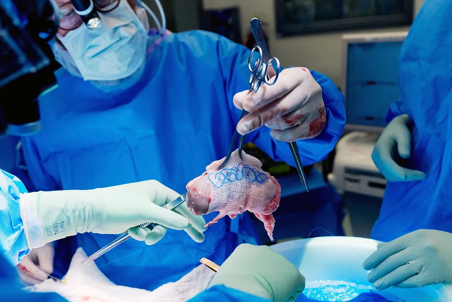


Three kinds of DNA evidence in one hospital test
Siriraj Hospital in Thailand has introduced a brain tumour testing service that combines three kinds of molecular evidence in a single process, reducing the stated testing cost from about 55,000 baht to 27,000 baht and shortening the wait for results from roughly 30 days to between one and five days.
Contents
- Three kinds of DNA evidence in one hospital test
- Why a tumour's appearance is only part of the diagnosis
- A Thai study changed six of 32 diagnoses
- How nanopore testing combines the clues
- Specific DNA losses help determine tumour type and grade
- From a research programme to a national sample service
- The milestones behind the service
- What comes next for patients and hospitals
- Key Points
The service, developed at Mahidol University's Faculty of Medicine Siriraj Hospital under the Genomics Thailand programme, examines gene mutations, changes in the number of copies of DNA and chemical markings called DNA methylation. Together, these clues help doctors identify a tumour more precisely and decide how to treat it.
The research team analysed 200 tumour samples from Thai patients using Oxford Nanopore technology, reading genetic information across the genome and examining methylation at more than 20 million positions. Researchers used those data to develop a tumour classification model suited to the Thai population.
Siriraj now accepts samples from hospitals throughout Thailand through its Advance Lab Service, part of the Siriraj Specimen Management Center. The technology is being used for Thai and foreign patients undergoing brain tumour surgery in the country.
The stated service turnaround is one to five days, while the project also sets a target of returning results within one day. At approximately 27,000 baht, testing would cost 28,000 baht less than the previous figure, a reduction of about 51%. For families facing surgery and treatment decisions, the practical change is access to a detailed molecular report inside Thailand, with less reliance on sending tissue abroad.
Why a tumour's appearance is only part of the diagnosis
Brain tumours can look similar under a microscope while having different genetic features. MRI and CT scans show doctors where a tumour is and how it affects nearby tissue. Examining a tissue sample adds information about its cells. Molecular testing supplies another layer: the biological features that help define which tumour a patient has.
The World Health Organization's 2021 classification of central nervous system tumours gives molecular findings an important role in identifying tumour types and grades. A grade describes biological behaviour, including how aggressively a tumour is likely to grow.
For patients, a more precise classification can change the expected course of the illness and inform treatment choices. The aim is to give pathologists, who diagnose disease by examining tissue, and treating doctors a fuller picture of the same tumour.
A Thai study changed six of 32 diagnoses
A glioma research report published in the Health Systems Research Institute's repository shows why these molecular details matter. Researchers examined samples from 32 patients with glioma, a group of brain tumours, using several methods to study DNA, RNA and methylation.
DNA methylation profiling changed the diagnosis in six of the 32 cases, or 19%. The report says those changes directly affected assessment of the likely course of disease and identification of appropriate treatment.
The researchers used whole exome sequencing, which examines the parts of DNA that contain instructions for making proteins, alongside RNA sequencing and methylation profiling. RNA helps cells put genetic instructions into action, so studying it can reveal changes that complement the DNA findings.
The 32 patient study and the 200 sample development programme address related parts of the diagnostic challenge. The former documents how detailed molecular analysis changed diagnoses; the latter supplied data for the integrated nanopore classification service.
How nanopore testing combines the clues
Nanopore technology reads DNA as it passes through extremely small pores. Changes in an electrical signal help identify the DNA sequence and chemical modifications attached to it. Its ability to examine long stretches of DNA helps researchers study several features within one testing process.
Siriraj's system brings three groups of markers into a report for doctors:
- Gene mutations: changes in the DNA sequence that can help identify a tumour's biological features.
- Copy number variation, or CNV: gains or losses of sections of DNA, which can help classify a tumour and determine its grade.
- DNA methylation: chemical markings on DNA whose patterns can help distinguish tumour types.
DNA methylation can be understood as a set of chemical labels on genetic material. Different tumours can carry different patterns of these labels, giving doctors another way to identify them when other findings leave uncertainty.
The Thai team also developed a tool called GLIMMERS to bring the findings together and prepare reports for clinicians. This matters because sequencing produces large amounts of data. Doctors need the relevant findings organised into information they can use alongside scans, tissue examination and the patient's condition.
Specific DNA losses help determine tumour type and grade
The Health Systems Research Institute report describes an earlier nanopore method for detecting two changes important to the WHO's 2021 classification of gliomas with an IDH mutation. IDH refers to genes whose mutation status helps divide gliomas into biologically different groups.
One change is 1p/19q co-deletion, the combined loss of particular parts of chromosomes 1 and 19. The other is CDKN2A/B homozygous deletion, meaning both copies of that gene region are missing. These findings help doctors assign the correct tumour category and grade.
The researchers reported close agreement between CNV findings obtained through nanopore sequencing and those obtained through methylation and exome analysis, with lower costs and shorter processing times. The nanopore CNV work was published in Brain Pathology in August 2023. The full citation is Wongsurawat et al., "Exploiting nanopore sequencing for characterization and grading of IDH-mutant gliomas", Brain Pathology 34(1):e13203, published online August 13, 2023; DOI: 10.1111/bpa.13203.
The wider study also identified gene fusions, in which parts of different genes become joined: FGFR3-TACC3 in two cases and EGFR-SEPT14 in one. Researchers explored immune cells in 10 glioma samples using digital pathology, which analyses digitised tissue images. They described that work as exploratory and called for validation in larger groups.
From a research programme to a national sample service
Dr Thidathip Wongsurawat of the Siriraj Genomics Center leads the integrated nanopore project, supported by the Health Systems Research Institute under Genomics Thailand. The work brings together doctors and specialists in bioinformatics, the use of computing to interpret biological data.
The system accepts fresh tissue and formalin fixed, paraffin embedded tissue, commonly called FFPE. In FFPE processing, tissue is preserved with a chemical fixative and embedded in wax, a routine way for pathology departments to store samples. Supporting this format allows the service to use material hospitals already handle.
Hospitals across Thailand can send samples to Siriraj's Advance Lab Service. The research network includes Mahidol, Chulalongkorn, Chiang Mai, Khon Kaen and Prince of Songkla universities, alongside a pathology institute and Nhealth within the Bangkok Hospital network.
The network connects university hospitals in several regions with public and private participants. For hospitals referring samples, the central service provides access to specialised analysis without requiring each institution to establish the entire laboratory and computing process itself.
The milestones behind the service
The development spans changes in international tumour classification, Thai genomic research and the introduction of hospital testing. The main dated milestones are:
- 2021: The WHO classification of central nervous system tumours gives molecular markers an important role in tumour identification and grading.
- August 2023: The Thai team's nanopore research on detecting DNA copy number changes is published in Brain Pathology.
- 2026: The integrated brain tumour classification project wins a National Innovation Award in the social category for public sector and nonprofit organisations.
- November 18, 2026: Details of the service describe the 200 sample development programme, nationwide sample acceptance and a turnaround of one to five days.
Thailand's National Innovation Agency, a public organisation, awarded the project the top prize in its category. The recognised work combines multiple biomarkers through nanopore technology, carrying a research approach into a service available within the hospital system.
What comes next for patients and hospitals
The research team has filed an international patent application under the Patent Cooperation Treaty for a method titled "Method for Brain Tumor Classification based on The Integrative Analysis of Multiple Biomarkers". The application covers the approach of combining different biological markers to classify brain tumours.
The next phase includes broader testing with the hospital network and research into using artificial intelligence to help interpret clinical findings according to WHO criteria. The team also plans to develop testing kits meeting ISO standards, with the aim of expanding use overseas and generating income for Thailand.
No timetable has been given for the planned ISO standard testing kits or the introduction of AI assisted clinical interpretation. These remain development goals alongside the existing sample testing service.
For patients now, the main benefit is a shorter route to molecular information that can guide a diagnosis. Surgery, pathology and treatment planning remain part of their care. The new service puts three major groups of molecular findings into one report, giving doctors more information with which to choose the next step.
Key Points
- Siriraj combines gene mutations, DNA copy number changes and methylation in one nanopore testing process.
- The stated cost falls from about 55,000 baht to 27,000 baht, roughly 51% less.
- Results are described as available within one to five days, with a target of one day.
- The classification programme analysed 200 Thai tumour samples and more than 20 million methylation positions.
- A related study found that methylation profiling changed six of 32 glioma diagnoses.
- Siriraj accepts tissue samples from hospitals nationwide; wider network testing, ISO standard kits and AI research are planned.