
Tiny structures inside a Brazilian dinosaur bone
Microscopic structures measuring about 23 micrometers long and 20 micrometers wide, together with chemical traces of a molecule used in blood, have been found inside the leg bones of a dinosaur that lived in Brazil about 115 million years ago. Researchers interpret the structures as mineralized red blood cells, although an independent specialist disputes that identification.
Contents
- Tiny structures inside a Brazilian dinosaur bone
- A young predator with an unusual history
- From a gypsum mine to a molecular study
- How researchers connected shape with chemistry
- A manganese mineral may explain the preservation
- Why an independent specialist questions the cells
- What the comparison with bird blood adds
- Collagen traces add another piece of ancient biology
- Small museum fragments could hold more answers
- Key Points
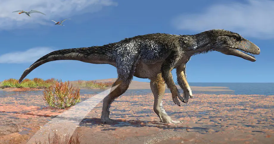
The study, published on November 2, 2026, in Proceedings of the National Academy of Sciences, examined Aratasaurus museunacionali, a predator that walked on two legs during the Early Cretaceous. The fossil came from a gypsum mine in Ceará, northeastern Brazil. Its surviving bones include parts of the right thigh, lower leg and foot.
A team led by Kliti Grice, an organic and isotope geochemist at Curtin University in Perth, Australia, detected heme B and a chemically altered relative called 3,8-diethyl heme B. Heme is the component containing iron that helps hemoglobin, the main oxygen transport protein in red blood cells, do its work.
The researchers place particular weight on finding the chemistry within tiny structures inside the bone's vascular canals, the channels that once carried blood vessels. Earlier studies had identified possible blood cell shapes in dinosaur fossils. This team argues that the combination of location, mineral preservation and chemical evidence makes its case stronger.
Grice, the study's lead author, described the discovery in terms of traces surviving through geological time:
We have not found a vial of dinosaur blood. What we are seeing is a mineral and molecular echo of ancient biology, and that is scientifically much more interesting.
A young predator with an unusual history
Aratasaurus was about 3.1 meters long and is estimated to have weighed roughly 35 kilograms. The individual studied was probably about four years old when it died, making it a juvenile or young adult that may still have been growing.
The fossil came from the Mina Pedra Branca site in the Romualdo Formation, part of the Chapada do Araripe region around the meeting point of Ceará, Pernambuco and Piauí. The formation is known for unusually well preserved fossils.
Alexander Kellner, a study coauthor and paleontologist at Brazil's National Museum, described the animal's remains as having reached the bottom of an ancient lake. Much of the skeleton was lost, but the surviving leg material eventually supplied both anatomical information and samples for chemical analysis.
The specimen also survived a much more recent disaster. It was undergoing preparation at the National Museum when fire devastated the institution in 2018. According to Kellner, the area holding the fossil escaped the flames.
From a gypsum mine to a molecular study
The discovery follows nearly two decades of work involving Brazilian and international researchers. Small fragments left over from preparing the fossil became valuable material for instruments capable of detecting extremely small quantities of organic compounds.
- 2008: The fossil was discovered in a gypsum mine in Ceará and taken to the Plácido Cidade Nuvens Museum of Paleontology in Santana do Cariri.
- 2016: The specimen was sent to the National Museum in Rio de Janeiro for further study.
- 2018: The fossil survived the National Museum fire.
- 2020: A team led by Juliana Sayão described Aratasaurus museunacionali as a new species.
- November 2, 2026: The study of its microscopic structures and blood related chemistry was published in PNAS.
Brazilian contributors to the new research included Renan Bantim and Antônio Saraiva of the Regional University of Cariri, and Sayão and Kellner of the Federal University of Rio de Janeiro's National Museum. Researchers from institutions in Germany, Japan and the United States also participated.
How researchers connected shape with chemistry
The team combined microscopy, mineral analysis and molecular chemistry. Each method addressed a different part of the puzzle: what the structures looked like, what preserved them and which organic compounds remained.
High performance liquid chromatography separated compounds extracted from the fossil. Mass spectrometry then helped identify them through their molecular masses and chemical patterns. The researchers compared measurements, including absorption of ultraviolet and visible light, with authentic modern standards.
Raman spectroscopy and electron microscopy supplied another layer of evidence. Raman analysis uses the way light interacts with a material to reveal its chemistry, while electron microscopes show details too small for ordinary light microscopes. Together, these methods helped locate heme related signals within the microscopic structures.
Grice said the value lay in connecting several observations in the same physical setting. A blood related molecule in an extract offers one clue; placing that chemistry inside a particular structure within a former blood vessel gives researchers a more detailed explanation to test.
The team also examined possible contamination and concluded that surrounding microorganisms or plants were unlikely sources of the detected heme. That assessment matters because fossil bones spend millions of years in contact with sediment, water and organisms that can introduce their own organic material.
A manganese mineral may explain the preservation
Hausmannite, a naturally occurring manganese oxide mineral, was associated with the structures identified as red blood cells. The researchers propose that mineral formation early in the fossilization process helped stabilize or copy the cells before decomposition destroyed their shapes.
Rapid burial may have helped too. The team's explanation involves an environment with little oxygen and microbial mats, thin layers of microorganisms living together on a surface. Such conditions could have slowed decay while creating opportunities for mineral preservation.
The proposed sequence connects the burial environment with the microscopic evidence: sediment covered the remains, unusual local chemistry encouraged mineral formation, and some organic compounds survived alongside the resulting structures. The timing and chemical steps remain a proposed explanation for the fossil's exceptional condition.
Heme is much smaller than an entire hemoglobin protein. Finding heme and its altered products therefore gives researchers access to a surviving chemical component of the animal's biology. The material recovered consists of mineralized structures and molecular traces rather than preserved liquid blood.
Why an independent specialist questions the cells
Mary Schweitzer, a molecular paleontologist at North Carolina State University who was not involved in the study, challenges the interpretation of the microscopic structures. Her objections concern their shape, size and lack of a visible feature expected in red blood cells from animals such as birds and reptiles.
Mature human red blood cells lose their nuclei, the compartments that hold genetic material. Birds and reptiles retain nuclei in their red blood cells, usually producing a noticeable bulge. Schweitzer said the fossil structures appear more concave and lack that bulge.
Schweitzer also considers the structures smaller than most red blood cells from animals outside the mammal group. Describing her objection to their appearance, she said:
morphological similarity is not met.
The study's authors base their interpretation on the combined evidence: numerous structures inside vascular canals, mineralization and heme related chemical signals associated with those structures. Schweitzer accepts that molecules originating within organisms can survive over geological time and welcomes the use of new molecular techniques, while remaining unconvinced that these particular structures are red blood cells.
The central disagreement is therefore about the identity of the structures. The authors see preserved cellular remains supported by chemistry; Schweitzer finds their visible anatomy a poor match for the cells expected in this dinosaur.
What the comparison with bird blood adds
The fossil structures averaged 22.97 micrometers in length and 19.54 micrometers in width. A micrometer is one thousandth of a millimeter, so these objects are far too small to inspect with the unaided eye.
The comparison presented with the research gives modern bird red blood cells a width of about 7.5 to 13 micrometers. Using those figures, the fossil structures are about 1.5 to 2.6 times as wide. They were described as almost spherical, while modern bird red blood cells are more visibly elongated.
The comparison with birds and Schweitzer's comparison with a broader range of animals lead to different assessments of size. Her criticism also includes the concave appearance and missing nuclear bulge, features that size measurements alone cannot resolve.
Cell dimensions interest researchers because oxygen transport is tied to an animal's energy needs. The team hopes molecular studies could eventually help investigate blood evolution, metabolism and activity in extinct animals. Direct estimates of Aratasaurus body temperature or activity levels were not presented.
Collagen traces add another piece of ancient biology
The fossil also yielded amino acids associated with collagen, the structural protein abundant in bone and other connective tissues. Amino acids are the small chemical units from which proteins are built.
Those findings widen the investigation beyond blood chemistry. The bone preserved evidence associated with its supporting tissue as well as compounds linked to oxygen transport, giving researchers more than one category of ancient biological material to examine.
No intact ancient collagen protein or dinosaur DNA was recovered. Grice explained that large molecules carrying complex biological information have different preservation properties from small molecules and chemically altered remnants.
The oldest reported DNA cited in connection with the discovery comes from Greenland sediment dating to about two million years ago. The Aratasaurus fossil is about 115 million years old. Work on it concerns the survival of selected chemical traces across that much longer interval.
Grice said the research encourages scientists to keep investigating how long biological information can persist:
What the work does encourage us to do is keep testing the limits of molecular preservation rather than assuming that all biological information must disappear after a particular length of time.
Small museum fragments could hold more answers
The Aratasaurus analysis relied on tiny pieces left from fossil preparation. That detail gives the discovery practical relevance for museums, where fragments retained during earlier work may contain chemistry that older methods could not detect.
Some molecular tests require grinding or otherwise consuming a sample, making specimen selection important. Residual fragments can provide material for such analysis while keeping the main fossil available for anatomical research and display.
Further work could test whether similar combinations of vascular structures, minerals and heme chemistry occur in other specimens. Comparisons across fossils would also help researchers investigate the preservation process and address disagreements about cell identification.
Martin Van Kranendonk, director of Curtin University's School of Earth and Planetary Sciences, has also pointed to possible applications in astrobiology, the study of life in the universe. Understanding which biological traces survive hostile conditions on Earth can inform the search for traces of ancient life elsewhere.
For paleontology, the immediate opportunity is closer to hand: examining familiar collections with instruments that connect a fossil's microscopic anatomy to its chemistry. The Brazilian specimen shows why a few fragments from an incomplete leg can deserve another look.
Key Points
- A study published on November 2, 2026, identified heme related compounds in a dinosaur fossil about 115 million years old.
- The fossil belonged to a young Aratasaurus museunacionali from Ceará, Brazil, measuring about 3.1 meters long.
- Researchers interpret microscopic structures inside its bone's vascular canals as mineralized red blood cells.
- Independent paleontologist Mary Schweitzer disputes that identification, citing their shape, size and lack of a nuclear bulge.
- Hausmannite mineralization and rapid burial in an environment with little oxygen may have helped preserve the structures and chemistry.
- The team also detected amino acids associated with collagen. No intact ancient protein or dinosaur DNA was recovered.
- The findings create opportunities to investigate molecular traces in fragments already held by museums.