
A protein disposal pathway offers a new research target
Researchers at Nagoya University and collaborating institutions have identified a pathway that helps cells break down TDP-43, a protein that accumulates abnormally in many people with amyotrophic lateral sclerosis, or ALS. The finding points to a possible way to tackle one of the processes linked to the death of nerve cells that control movement.
Contents
The study, titled TBK1 eliminates aggregation-prone monomeric TDP-43 through an IFNβ-immunoproteasome pathway in ALS models, was published open access in EMBO Reports on October 5, 2026, with the DOI 10.1038/s44319-026-00953-8. Its authors include Shohei Sakai, Kotaro Oiwa, Yohei Iguchi, Seiji Watanabe, Masahisa Katsuno and Koji Yamanaka.
The pathway involves a protein called TBK1, an immune messenger called interferon beta, or IFNβ, and cellular machinery known as the immunoproteasome. The researchers found that interaction with TDP-43 activates TBK1. Activated TBK1 then acts through IFNβ to induce the immunoproteasome, which breaks down and removes TDP-43.
The team suggests that this disposal system may function poorly in people with ALS, allowing the protein to build up. Understanding where that process fails could help researchers identify ways to support the cell's own ability to clear harmful material.
A separate Nagoya University study, published on January 10, 2024, identified another route involving an enzyme called IKKβ. That work showed how the enzyme could promote the breakdown of abnormal TDP-43 in the cell's cytoplasm while leaving normal TDP-43 in the nucleus unaffected.
The two findings give researchers different routes to investigate around the same problem: how to remove harmful TDP-43 while preserving the protein's normal work. The TBK1 discovery concerns an immune signaling pathway that activates protein disposal machinery. The IKKβ study describes a chemical modification that helps direct TDP-43 toward breakdown.
For patients and families, the immediate significance is a new research direction. Whether either pathway can be turned into a safe treatment that slows ALS in people, and when human trials might begin, has not been established.
Why TDP-43 matters in ALS
ALS is a progressive disease in which motor neurons, the nerve cells that send instructions to muscles, die. As those instructions disappear, muscles weaken and shrink. Movement becomes more difficult, and the disease can eventually affect the muscles needed for swallowing and breathing.
TDP-43 normally works mainly inside the nucleus, the compartment that holds a cell's genetic material. It helps manage RNA, the molecules involved in carrying and processing genetic instructions. Cells therefore need a working supply of this protein.
In ALS, TDP-43 can leave the nucleus and accumulate in the cytoplasm, the surrounding part of the cell. There, it can form aggregates, or clumps. These abnormal deposits are considered a major contributor to motor neuron damage.
Nagoya University's 2024 research announcement states that sporadic ALS accounts for more than 90% of cases. Sporadic means the disease occurs without an apparent inherited family pattern. The university says the cause of this form remains unknown and that treatments do not sufficiently suppress its progression.
The treatment challenge is therefore more precise than simply reducing the amount of TDP-43 everywhere. Researchers want to limit harmful accumulation while protecting the normal protein that cells rely on.
How TBK1 activates the disposal process
TBK1 acts near the beginning of the newly described pathway. The researchers found that it becomes active when it interacts with TDP-43, connecting the presence of the protein to a response that can help remove it.
Activated TBK1 acts through IFNβ, a cytokine. Cytokines are messenger proteins that allow cells to communicate and coordinate responses. In this pathway, IFNβ helps induce the immunoproteasome.
A proteasome is a molecular machine that breaks proteins into smaller pieces. Cells use these machines to dispose of proteins they no longer need or that have become damaged. The immunoproteasome is a specialized form associated with immune responses.
The sequence described by the researchers links those functions together: TDP-43 interacts with TBK1, TBK1 becomes active, IFNβ carries the signal, and the immunoproteasome helps degrade TDP-43. A protein associated with harmful buildup can therefore also participate in activating a pathway for its removal.
The study traced this signaling through TBK1, STING, IRF3, IFNβ and JAK/STAT to the immunoproteasome. This route clears aggregation-prone monomeric TDP-43, the form consisting of individual protein molecules, independently of autophagy, another cellular disposal process. "Here, we identify a TBK1-IFNβ-immunoproteasome axis as a novel autophagy-independent proteostatic pathway regulating TDP-43 clearance," the authors wrote.
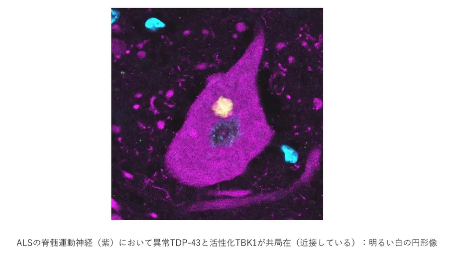
The work included experiments in neuronal cells and human lower motor neurons derived from induced pluripotent stem cells, as well as examination of spinal motor neurons from people with ALS and experiments in SOD1G93A ALS model mice with one copy of Tbk1 deleted. IFNβ reduced monomeric TDP-43 in the stem cell-derived motor neurons. In patient tissue, IFNβ receptor expression was reduced in spinal motor neurons with TDP-43 pathology. In the mice, deletion of one Tbk1 copy impaired immunoproteasome induction and increased the accumulation of proteins tagged with ubiquitin in the spinal cord.
The researchers propose that inadequate operation of this system may contribute to abnormal TDP-43 accumulation in ALS. That gives future work a concrete focus: examining which part of the chain fails and whether its activity can be restored.
What the earlier IKKβ study found
Professor Masahisa Katsuno and lecturer Yohei Iguchi of Nagoya University's Graduate School of Medicine led the separate work on IKKβ, with Iguchi serving as first author. Their findings appeared in the Journal of Cell Biology on January 10, 2024.
The university's IKKβ research announcement describes an enzyme that selectively promotes the breakdown of TDP-43 in the cytoplasm. The paper is titled IκB kinase phosphorylates cytoplasmic TDP-43 and promotes its proteasome degradation, with the DOI 10.1083/jcb.202302048.
IKKβ is a kinase, a type of enzyme that attaches phosphate groups to proteins. This process, called phosphorylation, can change how a protein behaves, where it goes or how other cellular machinery handles it.
The team found that IKKβ modifies TDP-43 particularly at Ser92. This name identifies a serine, one of the protein's amino acid building blocks, at position 92. Modification at this site promoted the protein's breakdown through the proteasome.
The location of that modification matters. Abnormal TDP-43 in ALS was already known to carry excessive phosphate groups near its carboxyl end, one end of the protein. The researchers identified a role for phosphorylation elsewhere, especially at Ser92, closer to the opposite end.
Phosphorylation can have different effects depending on where it occurs. Here, the modification was associated with faster disposal of TDP-43, providing a specific molecular step for researchers to examine.
Preserving useful protein while reducing damage
IKKβ left normal TDP-43 functioning in the nucleus unaffected while directing pathological TDP-43 in the cytoplasm toward degradation, according to the university. That selectivity addresses a central difficulty in targeting a protein that is both necessary for normal cell activity and involved in disease.
The researchers also tested the effect in nerve cells from the mouse hippocampus, a brain region involved in learning and memory. They found that IKKβ reduced TDP-43 aggregation and lessened the damage associated with those aggregates.
The combination matters: fewer clumps were accompanied by less injury to nerve cells in the experiment. The work therefore examined a biological consequence of the protein buildup as well as the amount of accumulated protein.
The experiment used hippocampal neurons, while ALS chiefly damages motor neurons. The next research challenge is to understand how the protective effect operates in the cells most directly affected by ALS and how it might be sustained in a living organism.
What spinal cord tissue revealed
The IKKβ team developed an antibody designed to recognize TDP-43 phosphorylated at Ser92. Antibodies can be used as highly specific detection tools, allowing researchers to locate a particular protein or chemical modification in tissue under a microscope.
When the researchers applied this antibody to spinal cord tissue from people with ALS, some aggregates inside motor neurons were stained. This showed that the Ser92 modification identified in the experiments was also present in human disease tissue.
The university interpreted the finding as suggesting that a mechanism for actively breaking down TDP-43 may already operate to some extent in ALS. Its activity may be too weak to keep pace with the disease process.
The TBK1 finding raises a related possibility through a different pathway: cells may have machinery capable of clearing TDP-43, yet fail to activate or maintain it adequately. Both research directions place attention on the balance between protein accumulation and the cell's capacity to remove it.
Why immune signaling needs careful study
TBK1, IFNβ and the immunoproteasome connect the new disposal pathway to immune activity. The IKKβ study also involves an enzyme with established immune functions, although its reported action on TDP-43 follows a different route.
IKKβ normally forms a complex with another kinase, IKKα, and a supporting protein called NEMO. This complex regulates NF-κB, a signaling system involved in immune responses, inflammation and other cellular activities. The Nagoya team investigated a role for IKKβ in ALS that had not previously been studied.
These wider functions make precision an important practical concern. A treatment intended to improve TDP-43 clearance would need to account for the other jobs performed by the proteins it targets. Researchers would also need to determine how much activity is useful and which cells need it.
The studies identify concrete biological processes to investigate: activation of the TBK1 and IFNβ pathway, induction of the immunoproteasome, and IKKβ modification of TDP-43 at Ser92. For ALS research, the shared goal is to make protein disposal effective enough to protect vulnerable neurons while preserving normal cellular functions.
Key Points
- Nagoya University researchers identified a pathway linking TBK1, IFNβ and the immunoproteasome to TDP-43 removal.
- The team suggests that inadequate activity of this pathway may contribute to protein accumulation in ALS.
- A separate study published on January 10, 2024, found that IKKβ promotes TDP-43 breakdown by modifying it particularly at Ser92.
- IKKβ acted selectively on cytoplasmic TDP-43 while leaving normal nuclear TDP-43 unaffected.
- Experiments in mouse hippocampal neurons showed reduced protein aggregation and reduced cellular damage.
- Some aggregates in spinal cord tissue from people with ALS carried the Ser92 modification.
- Developing these discoveries into treatments requires further research on effectiveness, delivery and safety.