
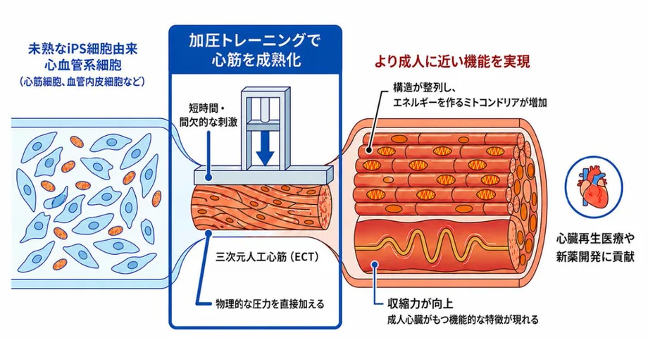
A short training routine changes artificial heart muscle
Japanese researchers have developed a way to make heart tissue grown from human iPS cells behave more like adult heart muscle: apply 50 kilopascals of pressure for one hour a day over three days, after an initial two weeks of culture. The trained tissue showed better organised muscle cells, stronger energy production and improved beating characteristics. When transplanted into rats with heart attack damage, it also improved heart function four weeks later.
Contents
- A short training routine changes artificial heart muscle
- What the researchers built
- Why three brief sessions worked best
- How the muscle became more capable
- A stronger beat, with a measurable limit
- Blood vessel cells help the whole tissue respond
- A second step builds vessel networks
- What happened after transplantation into rats
- Drug testing could come before clinical treatment
- Key Points
The collaboration involving RIKEN, Kyoto University, the University of Tokyo and Kansai Medical University announced the findings on July 24, 2026. The study appeared online in Stem Cell Reports on July 23, or July 24 in Japan. Its central finding is that brief pressure sessions worked better than continuous pressure or a longer training programme.
The advance addresses a persistent problem in regenerative medicine. Scientists can produce beating heart muscle from human induced pluripotent stem cells, usually called iPS cells, but the resulting muscle often resembles fetal tissue. Its contractions, energy use and responses to medicines differ from those of an adult heart.
The researchers moved their artificial tissue closer to an adult state across several measures, while identifying important limits. Its response to faster beating still peaked below the range reported for human heart muscle, and tissue made from a patient with an inherited heart condition responded less well. The immediate opportunity is a better research tool, with transplantation requiring further development and testing.
What the researchers built
Human iPS cells are cells reprogrammed into a state from which they can develop into many specialised cell types. For this study, the team used them to produce heart muscle cells and cells associated with blood vessels, then combined these with structural proteins, including collagen, to create engineered cardiac tissue, or ECT.
The result was a small, three dimensional tissue that could beat on its own. Heart muscle cells supplied its contracting machinery. Endothelial cells, which normally line blood vessels, and mural cells, which surround vessel walls, provided other parts of the tissue environment.
In one preparation described in the Stem Cell Reports study, heart muscle cells accounted for 39.12% of cells, endothelial cells for 26.49%, and mural cells for 12.81%. Those figures describe the tested preparation; the researchers also examined how differences in cell composition affected the response to pressure.
Hidetoshi Masumoto, a visiting senior researcher at RIKEN and the corresponding author, led the work with colleagues including RIKEN team director Wataru Kimura and University of Tokyo project associate professor Seitaro Nomura. Masumoto was a specially appointed professor in cardiovascular surgery at Kyoto University Hospital during the research and also holds a professorship at Kansai Medical University.
Why three brief sessions worked best
The team tested both the strength and timing of pressure. Continuous cyclic pressure at 10 or 50 kilopascals failed to increase the proportion of heart muscle cells. Intermittent sessions at 10, 50 and 100 kilopascals were then compared, with 50 kilopascals producing the preferred results.
Fifty kilopascals equals about 375 millimetres of mercury, the unit commonly used for blood pressure. The University of Tokyo Hospital announcement describes the experimental pressure as roughly 1,000 to 2,500 times the hydrostatic pressure during ordinary tissue culture. The treatment was applied to tissue in a laboratory pressure system.
Duration mattered just as much. Eight days of daily pressure sessions produced less benefit than three days and was associated with more apoptosis, the process by which cells undergo programmed death. Starting after two weeks of culture also produced a stronger response than starting after three weeks.
Masumoto described the discovery in Kyoto University's announcement. In a statement translated from Japanese, he said:
Through trial and error, we found, unexpectedly, that applying high pressure for only a short time was the most effective condition. The entire research team was surprised when the artificial heart muscle tissue changed substantially after just three hours of training.
The experimental sequence helps explain what those three hours involved:
- First two weeks: the engineered tissue was cultured before pressure training began.
- Next three days: tissue received 50 kilopascals of pressure for one hour each day, giving three hours of stimulation in total.
- After training: researchers examined structure, metabolism, gene activity and beating, and tested an additional culture step to encourage blood vessel networks.
- Four weeks after transplantation: rats with heart attack damage were assessed for heart function and survival of the implanted human tissue.
- July 23, 2026: the study was published online, with the Japanese announcement following on July 24.
How the muscle became more capable
Pressure training changed the tissue at several levels. Heart muscle cells became better aligned, and collagen within the tissue also showed improved alignment. An organised arrangement gives contracting cells a more coordinated structure in which to work.
Mitochondria, the tiny structures that produce much of a cell's usable energy, became more abundant. The trained tissue showed higher maximum oxygen consumption and greater production of ATP, the molecule cells use to power their activities. Isolated heart muscle cells were also larger and less round, changes consistent with maturation.
Calcium handling improved as well. Inside heart muscle cells, calcium acts as a signal that helps control when and how strongly the cells contract. The trained tissue showed larger calcium signals and faster calcium uptake and release during both spontaneous beating and electrical stimulation.
Researchers also found increased levels of SERCA2 and PLN, proteins involved in controlling calcium within cells. Taken together, the structural, energy and calcium findings offer a connected explanation for the improvement in beating: the tissue had a more organised muscle arrangement, more energy capacity and better control of its contraction signals.
A stronger beat, with a measurable limit
Adult heart muscle generally increases its contraction strength as its beating frequency rises within a certain range. Researchers call this a positive force-frequency relationship. The trained tissue acquired this characteristic, whereas untreated tissue tended to show declining movement as the pacing rate increased.
The study assessed contraction through video measurements of tissue movement using MUSCLEMOTION software. After training, movement increased when electrical pacing rose from 1 hertz to 1.25 hertz, equivalent to a rise from 60 to 75 beats per minute.
The response peaked at about 1.25 hertz. The paper cites a peak range of 2.0 to 2.5 hertz for human heart muscle, equivalent to 120 to 150 beats per minute. Full adult function therefore remains a development goal. The authors identify the conditions used to build the tissue, including its extracellular matrix, as possible limits. That matrix is the surrounding network of proteins that helps support and organise cells.
Cell composition also affected the outcome. Tissues showing a positive response contained an average of 63.5% heart muscle cells, compared with 45.9% in those showing a negative response, a difference of 17.6 percentage points. The researchers identified a proportion of 57.2% to 69.8% as favourable for pressure responsiveness in their analysis.
Blood vessel cells help the whole tissue respond
Endothelial cells emerged as an essential part of the method. When the researchers made tissue without these cells, pressure training produced little maturation. Their presence helped the muscle respond even before an internal blood vessel network had formed.
Endothelial cells normally encounter pressure and flowing blood in the body. They can sense physical forces and translate them into biological responses, a process called mechanotransduction. The experiments support a role for these cells in sensing pressure and promoting maturation throughout the engineered tissue.
Single cell RNA sequencing provided another view of the changes. This technique measures gene activity in individual cells, allowing researchers to see how different cell populations respond rather than averaging the whole tissue together.
Heart muscle cells in trained tissue showed increased activity in gene groups associated with mature muscle and reduced activity in developmental gene groups associated with immature muscle. The genetic findings matched the broader changes in structure, metabolism and beating.
A second step builds vessel networks
Pressure training alone left another task unfinished: the tissue contained endothelial cells, but an internal blood vessel network had not formed. The researchers therefore added dynamic culture after training, circulating the culture liquid around the tissue.
Circulation helps deliver oxygen and nutrients and creates a different physical environment from leaving tissue in still liquid. After this additional step, the team observed vessel networks inside the ECT and beating characteristics closer to those of adult heart muscle.
The combined approach separates two useful functions. Pressure sessions promoted muscle maturation, while the subsequent circulating culture supported vessel organisation. Both will matter as the researchers work toward larger implants, which need reliable access to oxygen and nutrients throughout their thickness.
What happened after transplantation into rats
The researchers implanted trained tissue into rats used as a model of myocardial infarction, or heart attack. Four weeks after transplantation, the animals showed improved heart function, including an increase in left ventricular ejection fraction.
The left ventricle is the chamber that pumps blood to the body. Ejection fraction measures the share of blood inside that chamber that is pushed out during each beat. An increase indicates improved pumping performance in this experiment.
Examination of the transplant sites found surviving human heart muscle tissue and the formation of a new muscle layer. These findings connect the laboratory maturation work with a possible use in repairing damaged hearts.
The next planned stages include making larger tissues, improving vessel formation and evaluating effectiveness and safety in large animals. The University of Tokyo announcement places possible clinical application within a research outlook of about five to ten years. No date for a human trial was given.
Drug testing could come before clinical treatment
The team is aiming for research use in drug development within a few years. More mature human heart tissue could give researchers a closer model for studying how medicines affect contraction, energy use and other heart functions, including unwanted effects.
Patient specific tissue offers another use. In this study, ECT made from iPS cells from a patient with hereditary dilated cardiomyopathy showed a limited response to pressure training. Dilated cardiomyopathy is a condition in which the heart enlarges and its pumping ability weakens.
The reduced response provides a feature researchers could investigate when studying the disease or testing treatment candidates. A pressure routine that produces different responses in healthy and diseased tissue may help reveal which parts of the maturation process are disrupted.
The practical attraction is the short stimulation schedule: three hours spread across three days, following the initial culture period. Consistent production will still require control over cell proportions, tissue structure and vessel development. Support for the work included RIKEN's Organoid Project, the Japan Society for the Promotion of Science grant JP22H03161, AMED's BINDS programme and the Novartis Global Scholars Program.
Key Points
- Pressure at 50 kilopascals for one hour daily over three days improved the maturation of heart tissue made from human iPS cells.
- The tissue showed better cell alignment, energy production, calcium handling and responses to faster pacing.
- Endothelial cells were essential to the maturation effect; circulating culture after training helped form vessel networks.
- Trained tissue improved heart function four weeks after transplantation into rats with heart attack damage.
- The tissue's frequency response remained below the adult human range, and tissue from a patient with inherited heart disease responded less well.
- Drug research use is targeted within a few years, while possible clinical application is viewed on a horizon of about five to ten years.