
A cell surface protein connects two routes of rubella infection
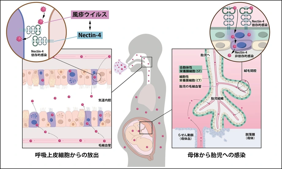
A Japanese research team has identified Nectin-4, a protein that normally helps cells stick together, as a major gateway used by rubella virus to enter human airway and placental cells. The discovery connects two central problems in rubella: how the virus reaches the respiratory tract for release and how it infects the boundary between a pregnant woman and her developing baby.
Contents
- A cell surface protein connects two routes of rubella infection
- Why the placental finding matters
- How researchers narrowed the search
- Adding, removing and restoring the receptor
- The virus attaches first, then moves inside
- Airway cells have an entrance side and an exit side
- A target at the maternal and fetal boundary
- What drug development would need to address
- From the preliminary paper to shared research data
- Key Points
The team includes researchers from the Japan Institute for Health Security (JIHS), Tohoku University, Osaka University and other Japanese institutions. Their Nature research paper describes experiments in which adding Nectin-4 made previously unsusceptible cells vulnerable to rubella, while removing or blocking the protein reduced infection.
The Nature publication record gives November 4, 2026, as the publication date; university announcements give November 5. The findings build on a preliminary paper posted in November 2025.
Nectin-4 is also a receptor for measles virus, despite measles and rubella belonging to different virus families. A receptor is a molecule on a cell that a virus can bind to and use during entry. Identifying it gives researchers a specific interaction to investigate when designing ways to interrupt infection.
Why the placental finding matters
Rubella spreads through respiratory droplets and contact. Its main symptoms include fever, a rash and swollen lymph nodes. Infection during early pregnancy can have much more serious consequences because the virus can pass from mother to fetus.
Congenital rubella syndrome, or CRS, can cause hearing loss, cataracts and heart disease. The new work helps explain how rubella infects a particular layer of the placenta, the organ that supports the developing baby and exchanges materials with the mother's blood.
Vaccines against rubella are already widely used. The research opens a different line of investigation: whether the interaction between the virus and Nectin-4 can become a target for future antiviral drugs or preventive treatments. The work remains at the laboratory stage.
How researchers narrowed the search
The researchers combined a search for proteins near virus particles with genetic tests of which proteins infection actually needed. Tohoku University's research announcement describes the use of viruslike particles, protein analysis and gene knockout screening to identify Nectin-4.
Viruslike particles reproduce useful features of a virus for experiments. In this study, the team used them with proximity biotin labelling, a technique that marks nearby proteins so those proteins can be collected and identified. Proteomics means studying many proteins together rather than testing them one at a time.
The team's November 2025 preprint, a preliminary version made public before journal publication, describes 89 proteins found specifically in the treated cells compared with untreated controls. Researchers then narrowed the candidates using information about their location on cells and their presence in susceptible cell types.
CRISPR–Cas9 supplied the next test. This gene editing system allowed researchers to disable selected genes and examine what happened to infection. After two rounds of rubella infection, both guide RNAs targeting NECTIN4 were enriched among surviving cells. Guide RNAs direct the editing machinery to a chosen gene; the result pointed to loss of Nectin-4 as a route to protection in those experiments.
Adding, removing and restoring the receptor
Nectin-4 passed several tests beyond the initial screen. Introducing the human protein into RajiSC cells supported growth of both a wild type rubella strain and a vaccine strain. Introducing another candidate, human MOG, did not support growth under the same experimental conditions, according to the preprint.
The team also removed the Nectin-4 gene from NCI-H358 cells, a human lung cancer cell line that naturally produces the protein. Infection fell. Restoring human Nectin-4 restored susceptibility, linking the change in infection to the receptor.
Antibodies directed against Nectin-4 supplied another way to interrupt the process. Blocking the receptor reduced infection in the experimental systems, including respiratory and placental models. Together, the genetic and antibody experiments gave the researchers several ways to test the same proposed entry route.
The inclusion of a vaccine strain helped establish the range of viruses able to use the receptor in cultured cells. The research examined their entry machinery rather than comparing vaccination outcomes in people.
The virus attaches first, then moves inside
The Nature paper describes entry as a sequence of events. Rubella first attaches to lipids, the fatty molecules in the cell membrane, through an interaction that depends on calcium. It then binds Nectin-4 and is taken into the cell.
Nectin-4 directly interacts with the virus's envelope glycoprotein heterodimer. In simpler terms, this is a pair of proteins, E2 and E1, on the virus's outer surface. Their interaction with the receptor helps the cell draw the virus inward.
The process is called receptor mediated endocytosis. The cell membrane folds around material at its surface and carries it into an internal compartment. The preprint's proposed model then describes how acidity inside that compartment triggers changes in the viral E1 protein, allowing the viral envelope to fuse with the compartment membrane and release viral contents.
This sequence gives researchers several steps to study: initial attachment, receptor binding, uptake and membrane fusion. The study's strongest new contribution is identifying Nectin-4 as a functional receptor and connecting it to infection in relevant human cell models.
Airway cells have an entrance side and an exit side
Human airway lining cells have different surfaces with different jobs. The apical surface faces the airway space, where air and mucus pass. The basolateral surface faces underlying tissue and neighbouring cells. Keeping those surfaces distinct allowed the team to test which direction rubella used.
In differentiated airway cultures, rubella entered preferentially from the basolateral side. Newly produced viruses were released mainly from the apical side, toward the airway space. Nectin-4 helped mediate entry from the tissue facing surface.
The preprint describes a useful comparison within the same experimental approach. Respiratory syncytial virus and human coronavirus 229E preferentially infected the cultures when delivered to the apical surface, whereas rubella preferentially infected them from the basolateral surface.
Blocking Nectin-4 in the rubella experiments reduced both infection and the later release of newly produced virus. The findings connect a receptor on one side of the airway lining with viral release from the other, helping explain a cellular route through which rubella can reach the respiratory tract for shedding.
A target at the maternal and fetal boundary
The placental experiments focused on syncytiotrophoblasts, cells that form the outermost layer of placental villi. Villi are small branching structures involved in exchange between mother and fetus. Their outer covering is positioned at the interface with maternal blood.
Nectin-4 was abundant in this outer layer. Researchers investigated its role using differentiated placental cells and trophoblast organoids, small structures grown in the laboratory that reproduce selected features of placental tissue.
Antibody blockade of Nectin-4 reduced rubella infection in these models. This gives researchers a molecular explanation for how the virus can infect a cell layer at the maternal and fetal boundary, an early step relevant to congenital infection.
The final Nature paper also describes infection of other trophoblast populations through pathways that do not depend on Nectin-4. The placenta therefore presents more than one possible entry route. The receptor is a major part of the mechanism in syncytiotrophoblasts, while other placental cells can support infection differently.
What drug development would need to address
Gunma University's research release identifies the interaction between rubella virus and Nectin-4 as a potential target for prevention or treatment. Its account also names the National Institute of Health Sciences, Institute of Science Tokyo and Yokohama City University among the collaborating institutions.
A future treatment based on this finding would aim to interrupt viral entry. Researchers would need to determine how to block the virus effectively while accounting for Nectin-4's normal role in helping cells attach to one another. Pregnancy would require particular attention to the safety of both mother and fetus.
The preprint identifies a major experimental limitation: the absence of suitable animal models for clinically symptomatic rubella. The team therefore relied on differentiated cell cultures to study the receptor's biological role. Establishing safety and benefit in a living body remains a separate stage of development.
The authors also point to a wider biological gap. Nectin-4 is present in a restricted range of tissues, while rubella can infect a broader range. They propose that additional receptors probably help the virus infect other tissues and organs. Which molecules perform those roles remains unresolved.
From the preliminary paper to shared research data
The research developed across several public milestones. The preliminary paper set out the receptor experiments in 2025, followed by the Nature publication and university announcements in November 2026.
- November 13, 2025: The preliminary study was posted on Research Square.
- November 4, 2026: The Nature publication record dates the journal paper to this day; university announcements give November 5.
- November 9, 2026: Tohoku University's dated announcement described the airway and placental findings.
- By November 11, 2026: The discovery was being reported publicly in Japan.
The Nature paper also identifies repositories for the experimental data. Proteomics data are deposited under ProteomeXchange identifier PXD067722 and jPOST accession JPST004036. CRISPR screening sequence files are listed under DDBJ accession DRA022489. These identifiers allow other researchers to locate the underlying datasets.
The discovery brings together receptor identification, direct binding experiments and tests in airway and placental models. Its immediate contribution is a more detailed explanation of rubella infection, alongside a defined target for future laboratory work on prevention and treatment.
Key Points
- Japanese researchers identified Nectin-4 as a major receptor used by rubella virus to enter human airway and placental cells.
- Adding the receptor increased susceptibility in previously unsusceptible cells; removing or blocking it reduced infection.
- Airway models showed preferential entry from the tissue facing side and release toward the airway space.
- Nectin-4 was abundant in syncytiotrophoblasts, the outer placental cell layer at the interface with maternal blood.
- Other placental cells supported entry through pathways independent of Nectin-4.
- The findings provide a target for treatment research, with further work needed on safety, other receptors and infection in living bodies.