
A chemical signal draws protective cells into damaged livers
Researchers at Kindai University in Japan have identified an immune mechanism that limits chronic liver damage in mice: a protein called CCL28 draws specialised antibody producing cells into the liver, where they help control inflammation and scarring.
Contents
- A chemical signal draws protective cells into damaged livers
- How the researchers tested liver protection
- What CCL28 tells immune cells to do
- Why these antibody cells behave differently
- How the cells acted on liver scarring
- Blocking the protective molecules worsened injury
- What the discovery means for treatment research
- Publication dates and study details
- Key Points
Mice lacking CCL28 developed more severe liver fibrosis, had higher blood levels of a marker of liver injury and accumulated fewer of these protective cells. The cells carried molecules called IL-10 and FasL, which helped restrain damage and act against cells responsible for producing scar tissue.
The study, published in Life Sciences, connects three parts of the process: a signal that attracts immune cells, a receptor that helps those cells respond, and molecules through which they regulate inflammation and scar formation. Its findings come from experiments in mice and laboratory cultures. Whether this mechanism can be used safely and effectively to treat people remains unknown.
The PubMed study record, maintained by the US National Library of Medicine, lists online publication on August 5, 2026. Kindai University says the final version appeared on October 15, followed by its announcement on October 16.
How the researchers tested liver protection
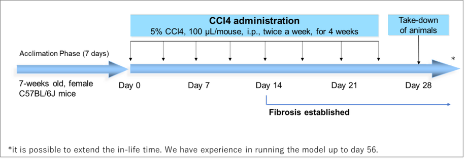
The Kindai team used carbon tetrachloride, known as CCl4, to cause repeated liver damage in mice. This chemical injury model allowed the researchers to examine how inflammation and fibrosis developed when CCL28 was present and when it was missing.
One group consisted of wild type mice, meaning animals without the targeted genetic deletion. The other consisted of CCL28 knockout mice, which had been genetically altered to lack CCL28. Comparing the groups helped the team investigate the protein's contribution to the liver's response to sustained injury.
Researchers examined liver tissue, measured gene activity and proteins, and used flow cytometry, a method that identifies and counts cells by their features. They also measured serum ALT, an enzyme that enters the blood when liver cells are injured.
The mice without CCL28 had higher ALT levels and more severe fibrosis. Their livers also contained fewer IgA antibody secreting cells, more activated hepatic stellate cells and fewer stellate cells undergoing programmed cell death. Together, these findings connected the missing immune signal with both greater injury and changes in the cells driving scar formation.
What CCL28 tells immune cells to do
CCL28 belongs to a family of proteins called chemokines. These proteins help direct the movement of immune cells, acting as chemical signals that guide cells towards particular tissues.
In the injured mouse livers, CCL28 expression increased in endothelial cells, which line blood vessels, and macrophages, immune cells involved in responding to tissue damage. The injured tissue was therefore producing more of a signal capable of attracting other immune cells.
The responding IgA cells carried CCR10, a receptor that recognises CCL28. A receptor is a protein through which a cell detects a particular signal. In this case, the CCL28 and CCR10 connection helped recruit cells with immune regulating activity into the liver.
Kindai's research announcement adds a detail about which cells responded: higher CCR10 expression on IgA cells was associated with higher expression of IL-10 and FasL. The cells better equipped to detect the recruitment signal also tended to carry more of the molecules involved in suppressing inflammation and controlling target cells.
Why these antibody cells behave differently
IgA is an antibody commonly associated with protection at mucosal surfaces, such as the lining of the intestine. Antibody secreting cells develop from B cells and produce antibodies that support the body's defences.
CCL28 was already known to help guide IgA cells to mucosal tissues. The Kindai study examined a different role involving a specialised group of IgA cells that can reduce immune activity during inflammation.
Many of the IgA cells that entered the damaged liver expressed IL-10, short for interleukin 10, and FasL, or Fas ligand. IL-10 helps suppress inflammatory responses. FasL can bind to a receptor called Fas on a target cell and trigger apoptosis, the controlled process through which a cell dies.
The protective effect therefore involved more than antibody production. These cells carried additional molecular tools that could quiet inflammation and influence the survival of other cells in the liver. Tracking those properties helped the researchers explain why fewer IgA cells accompanied worse damage in mice lacking CCL28.
How the cells acted on liver scarring
Hepatic stellate cells were a central target of the investigation. In a healthy liver, these cells store vitamin A. During injury, they can become activated and produce large amounts of collagen and other extracellular matrix proteins, the material that forms the structural framework around cells.
Too much of that material causes fibrosis, a buildup of scar tissue that makes the liver harder. Persistent liver damage can progress to cirrhosis, an advanced stage of scarring, and increase the risk of liver cancer.
The CCL28 knockout mice had more activated stellate cells and fewer stellate cells undergoing apoptosis. That combination helps explain the greater fibrosis: more cells were actively producing scar material, while fewer were being removed through programmed cell death.
Laboratory culture experiments tested this relationship more directly. B220 positive cells, a population identified using a cell marker and containing the immune regulating IgA cells, increased the killing of hepatic stellate cells. An antibody that blocked FasL reduced that effect.
The result connects the recruited immune cells with a specific action against the cells driving fibrosis. It also gives FasL a defined role in the experimental mechanism: helping trigger the death of stellate cells.
Blocking the protective molecules worsened injury
The team also interfered with IL-10 signalling and FasL activity in living mice. Treatment with an antibody against the IL-10 receptor or an antibody against FasL worsened chronic liver damage and fibrosis.
Blocking the IL-10 receptor prevents cells from responding normally to IL-10. Blocking FasL interrupts a route through which cells can trigger programmed death in a target. The worsening injury after these interventions supports a protective role for both molecular pathways in this model.
The experiments addressed different steps of the same process. Removing CCL28 disrupted the recruitment signal. Examining CCR10 identified how the IgA cells could respond to it. Blocking IL-10 signalling and FasL tested molecules used by the recruited cells, while the culture experiments examined their action against stellate cells.
For readers, the central finding is straightforward: some immune cells can protect an injured liver by reducing inflammation and helping remove cells that produce excessive scar tissue. CCL28 helps bring those protective cells to the damaged organ.
What the discovery means for treatment research
Kindai's team proposes that CCL28 or the specialised IgA cells could become starting points for new approaches to chronic liver disease. A treatment based on this mechanism would seek to control where protective cells gather or how they act after arriving.
Yuta Hara, a lecturer in Kindai University's Faculty of Pharmacy, described further work on chemokines and immune regulating IgA cells as a route towards developing new treatment strategies for liver disease. Professor Takashi Nakayama was the study's corresponding author.
The immediate research question is how a protective mechanism identified during chemical injury in mice relates to human liver disease. The study establishes an experimental role for CCL28, CCR10, IL-10 and FasL, while developing a medicine would require determining how to direct those activities towards the intended cells and tissues.
That targeting matters because the mechanism combines immune suppression with the ability to induce cell death. The proposed benefit depends on controlling damaging inflammation and scar producing cells. Kindai also points to possible research involving other immune related diseases, including cancer, as scientists investigate where these specialised IgA cells gather and what they do there.
Publication dates and study details
The paper is titled CCL28 ameliorates chronic liver damage associated with the recruitment of immunosuppressive IgA antibody-secreting cells in mice. It appears in Life Sciences, volume 403, as article 124615, with DOI 10.1016/j.lfs.2026.124615.
The publication sequence distinguishes the paper's initial online appearance from the final version described by the university:
- August 5, 2026: Online publication date listed in the PubMed record.
- October 15, 2026: Final publication date given by Kindai University.
- October 16, 2026: Kindai University announced the findings.
The research was conducted by a group in the Faculty of Pharmacy's chemotherapy laboratory at Kindai University in Higashiosaka, Osaka Prefecture. Its contribution is a connected explanation of how a recruitment signal, a specialised immune cell population and molecules acting on inflammation and stellate cells can limit chronic liver damage in an experimental setting.
Key Points
- CCL28 helped recruit immune regulating IgA cells into injured mouse livers through the receptor CCR10.
- Mice lacking CCL28 had fewer liver IgA cells, higher ALT levels and more severe fibrosis.
- The recruited cells expressed IL-10 and FasL, molecules involved in suppressing inflammation and triggering target cell death.
- Laboratory experiments connected FasL activity with the killing of hepatic stellate cells, which produce scar material.
- Blocking IL-10 signalling or FasL worsened liver damage and fibrosis in mice.
- The findings offer a direction for treatment research; their application to patients still requires investigation.